


Mohs Micrographic Surgery
Introduction
Mohs micrographic surgery, a surgical treatment for skin cancer that offers a very high rate of cure, was developed by Frederick E. Mohs, M.D., at the University of Wisconsin. The technique of Mohs surgery is time-consuming and requires highly specialized training and personnel. You were referred to us because Mohs micrographic surgery has been proven to be a highly successful form of treatment for your type of skin cancer.
The Mohs procedure will be performed by Joseph Giancola, M.D., a board certified dermatologist who is also a Fellow of the American College of Mohs Surgery (ACMS). Chosen through an extremely competitive review and selection process, fellows of the ACMS are required to complete an intensive 1-2 year post-residency fellowship training program. While any board certified dermatologist may perform Mohs surgery, only members of the ACMS have undergone rigorous fellowship training.
This information is designed to advise you about skin cancer as well as outline what you can expect on the day of your surgery and during the period after surgery. We have also included a section that outlines preventive measures you can take to decrease the possibility of developing skin cancers in the future. Please read all this information in its entirety before your surgery. If you have questions, call us at (602) 494-1817.
How Skin Cancer Forms
Cancer of the skin, like other cancers, is a disease of cells, which are tiny structures that make up all parts of the body. Although they differ in shape and function in various organs, all cells reproduce themselves by dividing. Normal growth and repair of tissue takes place in this orderly fashion.
When cell division is not orderly and controlled, abnormal growth occurs. Masses of tissue called tumors build up. Tumors can be benign or malignant. A malignant tumor is a cancer.
Benign tumors do not spread. But cancerous or malignant tumors invade and destroy surrounding normal tissue as they grow. Occasionally, cancer cells may break away from the tumor and spread (metastasize) through either the blood or lymphatic vessels to distant parts of the body where they form additional tumors.
Types of Skin Cancer
There are three primary forms of skin cancer: Basal cell carcinoma, squamous cell carcinoma and malignant melanoma.
Most cases of skin cancer are either basal cell carcinoma or squamous cell carcinoma. Of the two, basal cell carcinoma occurs more frequently and grows more slowly. It rarely spreads through the blood or lymphatic systems to distant parts of the body.
Squamous cell carcinoma is the second most common skin cancer. It is more serious, since it has a greater ability to spread internally to nearby lymph nodes and to other parts of the body.
Malignant melanoma may be life-threatening if not treated early. It usually appears as a brownish-black spot or bump in the skin that enlarges and sometimes bleeds. Sometimes melanomas arise in moles that have been present for many years.
Causes of Skin Cancer
Exposure to the sun is the leading cause of skin cancer, which commonly develops on the face, neck and arms as these are the most sun-exposed areas of the body. Fair-skinned people develop skin cancer more frequently than dark-skinned people. Cancers of the skin are most common in the southern United States.
Skin cancer also can be hereditary and occurs very frequently in certain ethnic groups, particularly those with fair complexion such as Northern Italians, Scandinavians and Celts (especially Irish and Anglo-Saxons). Other possible causes of skin cancer include X-rays, chronic injury and certain chemicals.
Signs of Skin Cancer
Although most skin growths are benign, any new growths on the skin or a sore that does not heal should be brought to your dermatologist’s attention.
Skin cancer has many different appearances. It may begin as a small, waxy lump that eventually bleeds and crusts; as a dry, scaly, red patch; or in several other ways. Although it may begin very small, skin cancer can grow to become very large.
Skin cancer may sometimes form from a noncancerous skin condition called actinic keratosis. These are red, rough patches of skin that develop as a result of sun damage and are commonly found on the face, neck or hands.
If the doctor thinks that a skin growth may be cancerous, a biopsy is performed. The whole area or a sample of the area is removed surgically and sent for examination under the microscope. The biopsy is used to confirm or rule out a diagnosis of cancer and to determine the type of any cancer found.
Treatment of Skin Cancer
Several treatment methods include surgical removal, or excision, with suturing (stitching); curettage and electrodessication, which is scraping and burning with an electric needle; radiotherapy (X-ray treatments); cryosurgery (freezing); and Mohs micrographic surgery. Many of these treatments have high cure rates, but Mohs micrographic surgery uniformly produces the highest success rate, especially for the most difficult tumors.
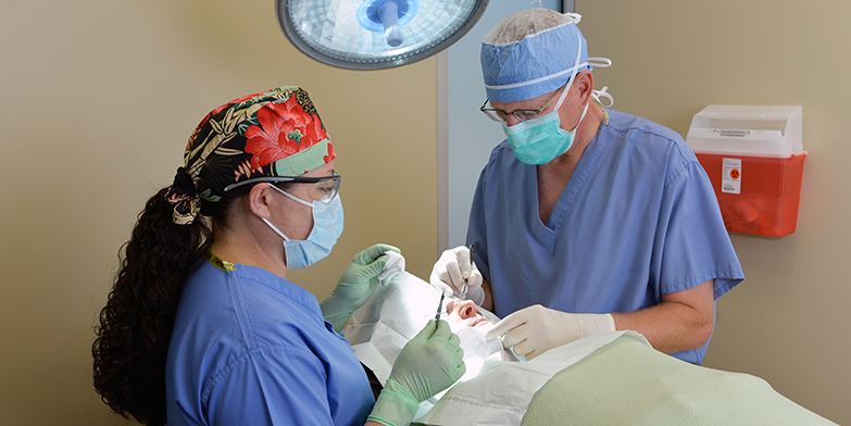
What You Should Know About Mohs Surgery
Mohs micrographic surgery is a specialized technique for the total removal of skin cancers. The procedure is done in several steps:
- A local anesthetic (usually lidocaine) is injected around the skin cancer.
- Surgical removal of the cancer is done in a specific fashion so that the entire undersurface and skin edges can be examined microscopically.
- The tissue is dyed and a corresponding map is drawn so that any tumor found microscopically can be located exactly on the patient.
- The surgeon then examines the slides under the microscope to look for cancer cells.
- Subsequent tissue is removed in a similar manner until no more cancer cells are found. In most cases, the surgery is done on an outpatient basis; however, in some cases, the doctor may recommend hospital admission. This is especially true for patients with large tumors, but admission is the exception rather than the rule.
How to Prepare for Surgery
No real preparation, other than a good night’s sleep, is required. Eat a light breakfast on the day of surgery. If you are currently taking medication, continue as usual unless directed otherwise by your physician. Eliminate aspirin or any medications containing aspirin, such as Bufferin or Anacin, for at least 7 days before surgery. This is because aspirin tends to prolong bleeding during the operation. Also, eliminate medications containing ibuprofen, commonly found in Advil, Motrin, and Aleve for at least 7 days before surgery. If you need a pain reliever, you may take acetaminophen, which is found in Tylenol.
It is best to wear a shirt that buttons down the front. No makeup or jewelry should be worn if surgery is to be performed on your face. Bring a good book or magazine with you, as you will spend a significant amount of time waiting while the microscopic slides are prepared and interpreted.
What to Expect on the Day of Surgery
Shortly after your arrival, you will be taken to one of the treatment rooms, where the tumor area will be numbed with a needle and local anesthetic.
The doctor will remove a thin layer of skin surrounding the cancer. After this has been done, any bleeding will be stopped with an electrocautery machine. You will then be bandaged and be able to return to the waiting room or you may stay in the treatment room. By this time, the removed tissue will be in our laboratory. There it is cut, dyed and made into microscopic slides. It usually takes 20-30 minutes for the layer of tissue to be removed and the bleeding to stop. However, it takes about 1 hour for the tissue to be prepared into microscopic slides for examination. During this time, you may chat with the person accompanying you, read a book or step out for fresh air.
If examination of the microscopic slides reveals that your tissue still contains tumor cells, the procedure will be repeated. Further tissue is removed only from the areas where tumor cells were found.
The goal is to remove all of the skin cancer while preserving the greatest amount of healthy tissue. However, skin cancers can grow deeply and develop roots extending beyond the area that you actually see. As a result, the final size of the surgical incision will be determined by the extent of the tumor.
The average number of surgical sessions required is 1-3. However, you may require more before your skin cancer is completely removed. Fortunately, this can usually be done in the course of a single day. When surgery is completed, a decision will be made as to the best way to manage your wound.
How Postoperative Wounds are Managed
Once it has been determined that your skin cancer has been completely removed, we will decide how best to manage your wound. In some instances, the wound should be allowed to heal by itself - this is called “healing by second intention.” In other cases, the wound needs to be repaired with stitches, or a skin graft or flap. The decision will depend on the size and location of your wounds. If a repair is needed, this usually can be done the same day or, in some cases, the following day. On rare occasions, you may be referred to another reconstructive surgical specialist.
If your wound is left to heal by itself, you will need daily bandage changes for 3-6 weeks. You will be given written instructions that describe how to change your bandages.
What to Expect After Surgery
Pain. Most patients do not have severe pain, but may experience slight discomfort. If this occurs, we suggest you take Tylenol every 4-6 hours as needed.
Bleeding. Occasionally, bleeding follows surgery. If this happens, do not become alarmed. Lie down and place steady, firm pressure over the wound as close as possible to the bleeding area. Apply the pressure continuously for 20 minutes (timed). Do not lift the bandage to check on the bleeding. If bleeding persists after 20 minutes of steady pressure, apply pressure for an additional 20 minutes. If bleeding still continues, call our office at (602) 494-1817 or go to the nearest hospital emergency room.
Swelling. Swelling is very common following surgery, particularly when it is performed near the eye. All wounds swell a little. Usually this is not a problem and the swelling diminishes as the wound heals. Ice packs and elevation may help reduce the swelling.
Drainage. All wounds will drain to some extent during the first 1-2 weeks. This is why frequent dressing changes are necessary.
Infection. Infection of the wound is unusual. However, if you see thick, foul-smelling fluid coming from the wound, or notice increasing redness, pain, warmth, or swelling call our office immediately. An antibiotic may be necessary.
Redness. All wounds will develop a halo of redness, which disappears gradually. If the area becomes extremely red and itchy, you may be allergic to either the ointment or the tape. Call our office if this condition develops.
Scarring. All surgeries leave a scar. However, your scar will improve and become less noticeable as time passes.
Stitches and skin grafts. If we close the wound with sutures (stitches) or place a skin graft, you should keep the area clean and follow the care instructions given to you until your next visit.
What to Expect After the Wound has Healed
You may experience some tightness, or drawing, as the wound heals. This is normal and usually lessens with time. Patients also commonly experience some itching after their wounds have healed. Rubbing a small amount of plain petroleum jelly on the scar often relieves this. Frequently, tumors involve nerves, and it may take up to a year, or even 2 years, before normal feeling returns to an area. Sometimes the area remains numb permanently. Only time will tell.
The scar tissue that grows over the wound contains many more blood vessels than the skin that was removed. This results in a red scar that may be sensitive to temperature changes. This sensitivity improves with time and the redness gradually fades.
Sometimes the scar is unacceptable to the patient and the patient’s family. If this is the case, surgical scar revision may be considered. However, we usually advise patients to wait 12 months before having the scar revision performed. This is because the scar continues to improve in appearance and becomes less conspicuous for up to 1 year after surgery.
Defense Against Future Skin Cancer
The most important thing you can do to lessen the possibility of developing future skin cancers is to protect your skin from further sun damage. This is easily achieved by the daily application of sunscreen to all exposed skin, including the tops of the ears. You also may wish to obtain further protection by wearing a broad-brimmed hat and appropriate protective clothing.
Apply the sunscreen 20 minutes before you go outdoors. It may wash off with water or heavy perspiration, so be sure to reapply a sunscreen after swimming or exercise, and use a sunscreen labeled “waterproof.”
A number of excellent sunscreens are available. Whatever sunscreen you choose, it should be broad-spectrum (block UVA/UVB) and have a sun-protection factor (SPF) of at least 30.
Followup Visits
Once your surgery is over, periodic follow up exams by your referring physician are important for at least 5 years. These visits can help with early detection and treatment of new skin cancers that may develop, as well as the rare recurrence of the skin cancer that was treated. If you notice any suspicious areas on your skin between visits, it is best to check with your doctor to see if a biopsy is needed.
For more information regarding Mohs surgery please visit the American College of Mohs Surgery at: www.mohscollege.org
Contact Us Today!
To learn more about our Cosmetic & Medical Dermatology Services, please contact us at 602-494-1817 to schedule an appointment!